Tuesday 8 June 2021

Blood Group || Rh-factor (+Ve/-Ve) & Erythroblastosis in hindi ||


Blood groups


There are 4 main blood groups (types of blood) – A, B, AB and O. Your blood group is determined by the genes you inherit from your parents.

Each group can be either Rh positive or Rh negative, which means in total there are 8 blood groups.

Antibodies and antigens

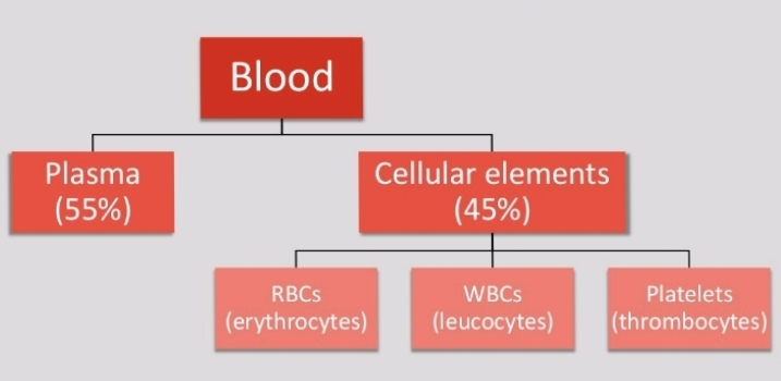
Blood is made up of red blood cells, white blood cells and platelets in a liquid called plasma. Your blood group is identified by antibodies and antigens in the blood.

Antibodies are proteins found in plasma. They're part of your body's natural defences. They recognise foreign substances, such as germs, and alert your immune system, which destroys them.

Antigens are protein molecules found on the surface of red blood cells.

The ABO system


There are 4 main blood groups defined by the ABO system:

blood group A – has A antigens on the red blood cells with anti-B antibodies in the plasma
blood group B – has B antigens with anti-A antibodies in the plasma
blood group O – has no antigens, but both anti-A and anti-B antibodies in the plasma
blood group AB – has both A and B antigens, but no antibodies
Blood group O is the most common blood group. Almost half of the UK population (48%) has blood group O.

Receiving blood from the wrong ABO group can be life threatening. For example, if someone with group B blood is given group A blood, their anti-A antibodies will attack the group A cells.

This is why group A blood must never be given to someone who has group B blood and vice versa.

As group O red blood cells do not have any A or B antigens, it can safely be given to any other group.


The Rh system

Red blood cells sometimes have another antigen, a protein known as the RhD antigen. If this is present, your blood group is RhD positive. If it's absent, your blood group is RhD negative.

This means you can be 1 of 8 blood groups:


A Rh positive (A+)
A Rh negative (A-)
B Rh positive (B+)
B Rh negative (B-)
O Rh positive (O+)
O Rh negative (O-)
AB Rh positive (AB+)
AB Rh negative (AB-)


Erythroblastosis

A hemolytic disease of the fetus and newborn that occurs when the immune system of an Rh-negative mother produces antibodies to an antigen in the blood of an Rh-positive fetus which cross the placenta and destroy fetal erythrocytes.

Sunday 6 June 2021

Blood Clotting Mechanism || Blood Coagulation in hindi ||

Blood Clotting


DEFINITION

Coagulation or clotting is defined as the process in which blood losses its fluid and becomes a jelly like mass few minutes after it is shed out ot collected in a container.


FACTORS INVOLVED IN BLOOD CLOTTING

Coagulation of blood occurs through a series of reactions due to the activation of a group of substances necessary for clotting are called clotting factors.


Thirteen clotting factors are identified.

1. Factor 1: Fibrinogen
2. Factor-II: Prothrombin
3. Factor III:Thromboplastin (Tissue Factor)
4. Factor-IV: Calcium
5. Factor-V: Labile Factor
6. Factor VI: Presence has not been proved
7. Factor VII: Stable factor
8. Factor VIII: Antihemophillic factor (Antihemophillic globulin)
9. Factor-IX:Christmas Factor
10. Factor-X: Stuart Prower factor
11. Factor-XI: Plasma Thromboplastin Antecedent
12. Factor-XII: Hageman factor (Contact factor)
13. Factor-XIII: Fibrin stabilizing Factor (Fibrinase)


STEPS OF BLOOD CLOTTING

In general blood clotting occurs in three stages.

1) Formation of Prothrombin activator

2) Conversion of prothrombin into thrombin

3) Conversion of fibrinogen into fibrin


STAGE 1: FORMATION OF PROTHROMBIN ACTIVATOR

Blood clotting commences with the formation of a substance called prothrombin activator, which converts prothrombin into thrombin.

Its formation is initiated by substances either within the blood or outside the blood.

Thus, formation of prothrombin activator occurs through two pathways:

1. Intrinsic Pathway

2. Extrinsic Pathway


STAGE 2: CONVERSION OF PROTHROMBIN INTO THROMBIN

Blood clot is all about thrombin formation. Once thrombin is formed definitely it leads to clot formation.

Prothrombin activator that is formed in intrinsic and extrinsic pathways converts prothrombin into thrombin. In the presence of calcium (factor IV). Once formed thrombin initiates the formation of more thrombin molecules.

The initially formed thrombin activates factor V.

Factor V in turn accelerates formation of both extrinsic and intrinsic prothrombin activator, which converts prothrombin into thrombin. The effect of thrombin is called positive feedback effect.


STAGE-3: FIBRINOGEN INTO FIBRIN

Sequence of events in stage 3 are as follows.

1. Thrombin converts inactive fibrinogen into activated fibrinogen .
The activatedfibrinogen is called fibrin monomer.

2. Fibrin monomer polymerizes with other monomer molecule and form loosely arranged strands of fibrin.

3. Later these loose strands are modified into dense and tight fibrin threads by fibrin stabilizing factor (Factor VIII) in the presence of calcium ions.

4. All the tight fibrin threads are aggregated to form a meshwork of stable clot.


Friday 4 June 2021

Human Blood || Plasma, RBC, WBC, And Platelet in hindi || #blood

Blood


Blood is a special type of fluid connective tissue derived from mesoderm.

The branch of science concerned with the study of blood, blood-forming tissues, and the disorders associated with them is called haematology.

haeme-blood and logos-study


PROPERTIES OF BLOOD


Colour : Bright red in arteries & dark red in veins

Mass : 8% of the body mass 

pH : Slightly alkaline (pH = 7.35-7.45)

Taste : Salty

Temperature : 38° C (100.4° F)

Viscosity : 3-4 times more viscous than water

Volume : 5-6 litre


                   Play Video


COMPOSITION OF BLOOD


Plasma

Plasma is a pale yellow coloured liquid component of a blood that holds the cellular elements of blood in suspension.


Constituents of plasma


Functions of plasma


Constituent.     Function

Water.        Absorbs, transports and releases heat

Albumins.         Osmotic balance

Globulins.        Defense mechanism

Fibrinogen.         Blood clotting

Electrolytic ions.       pH buffering


Red blood cells

Shape :- Circular biconcave non-nucleated

Size :- Diameter 7-8 ฮผm Thickness 2.5 ฮผm

Colour :- Red (haemoglobin pigment)

Count :- Adult male = 5.4 million RBCs/uL Adult female = 4.8 million RBCs/uL

Life Span :- 120 days


Erythropoiesis 

The production of RBCs is known as erythropoiesis.

Adult :- Red bone marrow of long bones (hip bone, breast bone & ribs)

Child (upto 5 year) : Bone marrow of all the bones

Foetus : Liver & spleen


Increase in number of RBCS is known as polycythemia

Decrease in number of RBCs is known as erythropenia


Functions of RBCs

Transport O₂ from lungs to tissues
Transport CO₂ from tissues to lungs

Normal blood contains 13-15 g of Hb per 100 ml of blood

One RBC contains about 250 million molecules of Hb

Each molecule of Hb carries four molecules of oxygen


White blood cells

Shape : Amoeboid nucleated

Size : 12-15 ฮผm

Colour : Colourless & translucent

Count : 5000-10000 WBCs/ฮผL

Life span : 10-13 days


Leucopoiesis

The production of WBCS is known as leucopoiesis.

Adult :- Liver, spleen, tonsils, bone marrow
Foetus :- Liver, spleen


Increase in number of WBCS is known as leucocytosis

Decrease in number of WBCS is known as leucopenia Pathological increase in number of WBCS is known as leukemia (blood cancer)


Types of WBCs


Platelets

Shape :- Circular biconvex non-nucleated

Size :- 2-4 ฮผm

Count :- 1,50,000 4,00,000 platelets/ฮผL

Life span :- 5-9 days

Function :- Blood clotting


Thrombopoiesis   


The production of platelets is known as thrombopoiesis.

Platelets are the fragments of large cells called megakaryocytes that remain in the bone marrow.

Increase in number of platelets is known as thrombocytosis

Decrease in number of platelets is known as thrombocytopenia


Wednesday 2 June 2021

Nervous tissue || Composition, structure, functions

Nervous tissue
Watch full video click here ๐Ÿ‘ˆ๐Ÿ‘‡


The most complex tissue in the human body is nerve tissue.

Formed by a network of more than 100 million nerve cells, assisted by many more glial cells.

Each neuron has, on an average, at least a thousand interconnection with other neurons forming a very complex nervous system.

Function

1. Regulates & controls body functions
2. Generates & transmits nerve impulses
3. Supports, insulates and protects impulse generating neurons.

Composition

The nerve tissue is composed of two elements:

1. The nerve cell or neuron 
2. The neuroglia


1- Nerve cell or Neuron

Nerve cells, also known as a neurons, are the active component of the nervous system.

Neurons communicate with each other as well as with other cells through electric signals (nerve impulses), which in turn allows effector organs to respond to the appropriate stimuli.

Function
Transmit electrical signals from sensory receptors and to effectors (muscles and glands) that control their activity.

Location:
 Brain, spinal cord and nerves.


2- Neuroglia or glial cells

Neuroglia, or glia, are the supportive cells for neurons.

They are non-neuronal cells in the central nervous system (brain and spinal cord) and the peripheral nervous system that do not produce electrical impulses.

They are involved in provision of nutrients, maintenance of a stable homeostatic environment, and the production of the myelin sheath.

Function:

Glia carry nutrients, speed repair, provide myelin for axons, support the blood-brain barrier, and may form their own communication network.

They are also involved in neurogenesis.

Location

Central nervous system (brain and spinal cord) and the peripheral nervous system

Moyamoya disease

Moyamoya disease is a disease in which certain arteries in the brain are constricted. Blood flow is blocked by constriction and blood clots...