Showing posts with label ummedsaini_. Show all posts

Friday, 4 March 2022

TRACHOMA

TRACHOMA


This is a chronic inflammation of the conjunctiva and the cornea of the eye. It is caused by an organism called Chlamydia trachomatis. Chlamydia trachomatis is also known to cause ophthalmia neonatorum, urethritis, cervicitis and salpingitis.

Trachoma is a major cause of blindness especially where water is scarce, such as among the pastoralist communities who inhabit the drier grasslands.

MODE OF TRANSMISSION

Trachoma is very common among communities living in dry areas where there is scarcity of water. Transmission of trachoma is by direct contact with the eye discharge of an infected person. Flies and fingers are important in the transmission of the disease.

CLINICAL PICTURE
Trachoma develops in 4 stages.

✓Stage 1: Early Trachoma
Initially the eyes are red and watery (as in ordinary conjunctivitis). After 30 or more days, follicles (small pinkish-grey lumps) form inside the upper eyelids.

Stage 2: Pannus Formation
Normally, the cornea has no blood capillaries on it. But during this stage, many tiny blood vessels are found to be growing towards the edge of the cornea. These tiny blood vessels which grow in the cornea are called pannus

Stage 3: Scarring of the Conjunctiva
After several years the follicles on the conjunctiva slowly begin to disappear leaving behind whitish scars on the conjunctiva. In the cornea, the small blood vessels degenerate. The vision becomes hazy.

Stage 4: Entropion and Trichiasis Formation
Due to this scarring, the scar tissue retracts (shortens), thereby causing the eyelids to become thick and to turn inwards. This is called entropion. As the thick, rough eyelids turn inwards, the eyelashes point inwards and rub against the cornea. This is called trichiasis. trichiasis adds to the damage already done to the eye and results in blindness

MANAGEMENT
• The drug of choice for the first three stages is 3% tetracycline topical eye ointment. 
• Stage four of the disease with entropion must be treated surgically.

PREVENTION & CONTROL
Trachoma is through supply of adequate water to the community. Regular bathing and washing of children's faces with water and soap should be encouraged. Reducing the fly population in the community through proper disposal of refuse and waste. Early treatment of infected individuals.

Tuesday, 8 June 2021

Blood Group || Rh-factor (+Ve/-Ve) & Erythroblastosis in hindi ||


Blood groups


There are 4 main blood groups (types of blood) – A, B, AB and O. Your blood group is determined by the genes you inherit from your parents.

Each group can be either Rh positive or Rh negative, which means in total there are 8 blood groups.

Antibodies and antigens

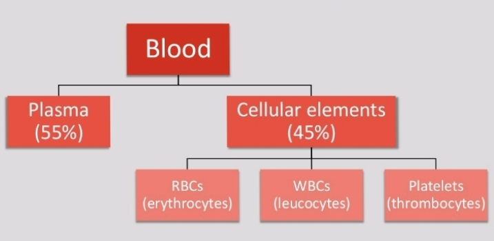
Blood is made up of red blood cells, white blood cells and platelets in a liquid called plasma. Your blood group is identified by antibodies and antigens in the blood.

Antibodies are proteins found in plasma. They're part of your body's natural defences. They recognise foreign substances, such as germs, and alert your immune system, which destroys them.

Antigens are protein molecules found on the surface of red blood cells.

The ABO system


There are 4 main blood groups defined by the ABO system:

blood group A – has A antigens on the red blood cells with anti-B antibodies in the plasma
blood group B – has B antigens with anti-A antibodies in the plasma
blood group O – has no antigens, but both anti-A and anti-B antibodies in the plasma
blood group AB – has both A and B antigens, but no antibodies
Blood group O is the most common blood group. Almost half of the UK population (48%) has blood group O.

Receiving blood from the wrong ABO group can be life threatening. For example, if someone with group B blood is given group A blood, their anti-A antibodies will attack the group A cells.

This is why group A blood must never be given to someone who has group B blood and vice versa.

As group O red blood cells do not have any A or B antigens, it can safely be given to any other group.


The Rh system

Red blood cells sometimes have another antigen, a protein known as the RhD antigen. If this is present, your blood group is RhD positive. If it's absent, your blood group is RhD negative.

This means you can be 1 of 8 blood groups:


A Rh positive (A+)
A Rh negative (A-)
B Rh positive (B+)
B Rh negative (B-)
O Rh positive (O+)
O Rh negative (O-)
AB Rh positive (AB+)
AB Rh negative (AB-)


Erythroblastosis

A hemolytic disease of the fetus and newborn that occurs when the immune system of an Rh-negative mother produces antibodies to an antigen in the blood of an Rh-positive fetus which cross the placenta and destroy fetal erythrocytes.

Sunday, 6 June 2021

Blood Clotting Mechanism || Blood Coagulation in hindi ||

Blood Clotting


DEFINITION

Coagulation or clotting is defined as the process in which blood losses its fluid and becomes a jelly like mass few minutes after it is shed out ot collected in a container.


FACTORS INVOLVED IN BLOOD CLOTTING

Coagulation of blood occurs through a series of reactions due to the activation of a group of substances necessary for clotting are called clotting factors.


Thirteen clotting factors are identified.

1. Factor 1: Fibrinogen
2. Factor-II: Prothrombin
3. Factor III:Thromboplastin (Tissue Factor)
4. Factor-IV: Calcium
5. Factor-V: Labile Factor
6. Factor VI: Presence has not been proved
7. Factor VII: Stable factor
8. Factor VIII: Antihemophillic factor (Antihemophillic globulin)
9. Factor-IX:Christmas Factor
10. Factor-X: Stuart Prower factor
11. Factor-XI: Plasma Thromboplastin Antecedent
12. Factor-XII: Hageman factor (Contact factor)
13. Factor-XIII: Fibrin stabilizing Factor (Fibrinase)


STEPS OF BLOOD CLOTTING

In general blood clotting occurs in three stages.

1) Formation of Prothrombin activator

2) Conversion of prothrombin into thrombin

3) Conversion of fibrinogen into fibrin


STAGE 1: FORMATION OF PROTHROMBIN ACTIVATOR

Blood clotting commences with the formation of a substance called prothrombin activator, which converts prothrombin into thrombin.

Its formation is initiated by substances either within the blood or outside the blood.

Thus, formation of prothrombin activator occurs through two pathways:

1. Intrinsic Pathway

2. Extrinsic Pathway


STAGE 2: CONVERSION OF PROTHROMBIN INTO THROMBIN

Blood clot is all about thrombin formation. Once thrombin is formed definitely it leads to clot formation.

Prothrombin activator that is formed in intrinsic and extrinsic pathways converts prothrombin into thrombin. In the presence of calcium (factor IV). Once formed thrombin initiates the formation of more thrombin molecules.

The initially formed thrombin activates factor V.

Factor V in turn accelerates formation of both extrinsic and intrinsic prothrombin activator, which converts prothrombin into thrombin. The effect of thrombin is called positive feedback effect.


STAGE-3: FIBRINOGEN INTO FIBRIN

Sequence of events in stage 3 are as follows.

1. Thrombin converts inactive fibrinogen into activated fibrinogen .
The activatedfibrinogen is called fibrin monomer.

2. Fibrin monomer polymerizes with other monomer molecule and form loosely arranged strands of fibrin.

3. Later these loose strands are modified into dense and tight fibrin threads by fibrin stabilizing factor (Factor VIII) in the presence of calcium ions.

4. All the tight fibrin threads are aggregated to form a meshwork of stable clot.


Friday, 4 June 2021

Human Blood || Plasma, RBC, WBC, And Platelet in hindi || #blood

Blood


Blood is a special type of fluid connective tissue derived from mesoderm.

The branch of science concerned with the study of blood, blood-forming tissues, and the disorders associated with them is called haematology.

haeme-blood and logos-study


PROPERTIES OF BLOOD


Colour : Bright red in arteries & dark red in veins

Mass : 8% of the body mass 

pH : Slightly alkaline (pH = 7.35-7.45)

Taste : Salty

Temperature : 38° C (100.4° F)

Viscosity : 3-4 times more viscous than water

Volume : 5-6 litre


                   Play Video


COMPOSITION OF BLOOD


Plasma

Plasma is a pale yellow coloured liquid component of a blood that holds the cellular elements of blood in suspension.


Constituents of plasma


Functions of plasma


Constituent.     Function

Water.        Absorbs, transports and releases heat

Albumins.         Osmotic balance

Globulins.        Defense mechanism

Fibrinogen.         Blood clotting

Electrolytic ions.       pH buffering


Red blood cells

Shape :- Circular biconcave non-nucleated

Size :- Diameter 7-8 ฮผm Thickness 2.5 ฮผm

Colour :- Red (haemoglobin pigment)

Count :- Adult male = 5.4 million RBCs/uL Adult female = 4.8 million RBCs/uL

Life Span :- 120 days


Erythropoiesis 

The production of RBCs is known as erythropoiesis.

Adult :- Red bone marrow of long bones (hip bone, breast bone & ribs)

Child (upto 5 year) : Bone marrow of all the bones

Foetus : Liver & spleen


Increase in number of RBCS is known as polycythemia

Decrease in number of RBCs is known as erythropenia


Functions of RBCs

Transport O₂ from lungs to tissues
Transport CO₂ from tissues to lungs

Normal blood contains 13-15 g of Hb per 100 ml of blood

One RBC contains about 250 million molecules of Hb

Each molecule of Hb carries four molecules of oxygen


White blood cells

Shape : Amoeboid nucleated

Size : 12-15 ฮผm

Colour : Colourless & translucent

Count : 5000-10000 WBCs/ฮผL

Life span : 10-13 days


Leucopoiesis

The production of WBCS is known as leucopoiesis.

Adult :- Liver, spleen, tonsils, bone marrow
Foetus :- Liver, spleen


Increase in number of WBCS is known as leucocytosis

Decrease in number of WBCS is known as leucopenia Pathological increase in number of WBCS is known as leukemia (blood cancer)


Types of WBCs


Platelets

Shape :- Circular biconvex non-nucleated

Size :- 2-4 ฮผm

Count :- 1,50,000 4,00,000 platelets/ฮผL

Life span :- 5-9 days

Function :- Blood clotting


Thrombopoiesis   


The production of platelets is known as thrombopoiesis.

Platelets are the fragments of large cells called megakaryocytes that remain in the bone marrow.

Increase in number of platelets is known as thrombocytosis

Decrease in number of platelets is known as thrombocytopenia


Wednesday, 2 June 2021

Nervous tissue || Composition, structure, functions

Nervous tissue
Watch full video click here ๐Ÿ‘ˆ๐Ÿ‘‡


The most complex tissue in the human body is nerve tissue.

Formed by a network of more than 100 million nerve cells, assisted by many more glial cells.

Each neuron has, on an average, at least a thousand interconnection with other neurons forming a very complex nervous system.

Function

1. Regulates & controls body functions
2. Generates & transmits nerve impulses
3. Supports, insulates and protects impulse generating neurons.

Composition

The nerve tissue is composed of two elements:

1. The nerve cell or neuron 
2. The neuroglia


1- Nerve cell or Neuron

Nerve cells, also known as a neurons, are the active component of the nervous system.

Neurons communicate with each other as well as with other cells through electric signals (nerve impulses), which in turn allows effector organs to respond to the appropriate stimuli.

Function
Transmit electrical signals from sensory receptors and to effectors (muscles and glands) that control their activity.

Location:
 Brain, spinal cord and nerves.


2- Neuroglia or glial cells

Neuroglia, or glia, are the supportive cells for neurons.

They are non-neuronal cells in the central nervous system (brain and spinal cord) and the peripheral nervous system that do not produce electrical impulses.

They are involved in provision of nutrients, maintenance of a stable homeostatic environment, and the production of the myelin sheath.

Function:

Glia carry nutrients, speed repair, provide myelin for axons, support the blood-brain barrier, and may form their own communication network.

They are also involved in neurogenesis.

Location

Central nervous system (brain and spinal cord) and the peripheral nervous system

Monday, 31 May 2021

Muscle tissue in hindi || Types, Function, Structure

 Muscle Tissue

Watch full video


Muscle Fibres เคธे Nerves, connected เคนोเคคे เคนैं।

'เคฎเคจुเคท्เคฏ เค•े เคถเคฐीเคฐ เคฎें 40 เคช्เคฐเคคिเคถเคค เคญाเค— Muscles เค•ा เคนोเคคा เคนै ।

Bone > Muscle >Blood

Structure, Function เคคเคฅा Location เค•े เค†เคงाเคฐ เคชเคฐ Muscle tissue เค•ो เคคीเคจ เคช्เคฐเค•ाเคฐ เค•े เคนोเคคे เคนैं ।


Classification

1. Striated muscle (เคฐेเค–ीเคฏ เคชेเคถी)

2. Smooth muscle (เคšिเค•เคจी เคชेเคถी)

3. Cardiac muscle (เค•ाเคฐ्เคกिเค• เคชेเคถी)


1 Striated muscle (เคฐेเค–ीเคฏ เคชेเคถी)

Striated muscles เค•ो skeletal or voluntary muscles เค•े เคจाเคฎ เคธे เคญी เคœाเคจเคคे เคนै।

เค•ुเค› Muscles เค•ो เคนเคฎ เค…เคชเคจी เคฎเคฐ्เคœी เคธे movement เค•เคฐा เคธเค•เคคे เคนैं เคœिเคธे voluntary muscles เค•เคนเคคे เคนैं।

เคฏเคน Long fibre เค•ा เคฌเคจा เคนोเคคा เคนै เคœिเคธเคฎे เคเค• เค•े เคฌाเคฆ เคเค• light เคคเคฅा dark band เคนोเคคा เคนै।

เค•ुเค› muscles เค…เคงिเค•เคคเคฐ เคนเคก्เคกिเคฏों เคธे เคœुเคก़े เคนोเคคे เคนै เคคเคฅा เคฌॉเคกी movement เคฎें เคธเคนाเคฏเค• เคนोเคคे เคนै เค‰เคธे Skeletal muscles เค•เคนเคคे เคนैं।


Striated muscle, Multinucleated เคนोเคคा เคนै।

Striated muscle, limb, body wall, face, neck เคฎें เคฆिเค–ाเคˆ เคชเคก़เคคा เคนै।

เคฏเคน tongue, pharynx, diaphragm เคฎें เค‰เคชเคธ्เคฅिเคค เคนोเคคा เคนै ।


Function :

Striated muscle เคฌเคนुเคค เคนी Powerful เคนोเคคा เคนै เคคเคฅा เคคेเคœी เคธे เคธिเค•ुเคก़เคคा เคนै।


เคฏเคน muscles เคฅเค• เคœाเคคा เคนै เคคเคฅा เค†เคฐाเคฎ เค•ी เคœเคฐूเคฐเคค เคชเคก़เคคी เคนै।


2. Smooth muscle (เคšिเค•เคจी เคชेเคถी)


เคœिเคธे เคนเคฎ เค…เคชเคจी เคฎเคฐ्เคœी เคธे Control เคฏा Movement เคจเคนीं เค•เคฐ เคธเค•เคคे เคนैं เคตเคน เค–ुเคฆ เคนी Contract เคฏा Relax เคนोเคคी เคนै।

Digestive system เคฎें Oesophagus เคธे เคฒेเค•เคฐ anus เคคเค• เค‡เคจ्เคนी muscle เค•ा เคฌเคจा เคนोเคคा เคนै।

Smooth muscle เคนเคก्เคกिเคฏों เคธे connected เคจเคนी เคนोเคคा เคนै।

Smooth muscle เคฎें เคœो cell เคชाเคฏा เคœाเคคा เคนै เค‰เคธเค•ा เคจाเคฎ myocytes เคนै เคœो fusiform เค†เค•ाเคฐ เค•ा เคนोเคคा เคนै เคช्เคฐเคค्เคฏेเค• muscle cell, plasma membrane เคธे เค˜िเคฐा เคฐเคนเคคा เคนै।


Function

Smooth muscle เค•े เค•ाเคฐเคฃ เค†เคนाเคฐ เคจเคฒी เคฎें เคญोเคœเคจ เค•ा Movement เคนोเคคा เคนै।

เค‡เคธเค•े เค•ाเคฐเคฃ เคนी Peristaltic movement เคนोเคคा เคนै เคœिเคธเคธे เค•ि เคนเคฎाเคฐा food เคนเคฎाเคฐे เคชेเคŸ เคคเค• เคชเคนुंเคšเคคा เคนै।

เค•ुเค› organ เคเคธे เคนोเคคे เคนैं เคœिเคจเคฎें smooth muscle เค•े เค•ाเคฐเคฃ เคนी extrusive movement (เคซैเคฒाเคต) produce เคนोเคคा เคนै เคœैเคธे urinary bladder, gall bladder or Uterus


3. Cardiac muscle (เค•ाเคฐ्เคกिเค• เคชेเคถी)

Cardiac muscle เคฆोเคจों smooth เคคเคฅा striated muscles เค•ा เค—ुเคฃ เคฆिเค–เคคा เค‡เคธ muscle เคฎें เคฌเคนुเคค เคธाเคฐे เคถाเค–ाเคं เคนोเคคी เคนैं เคœो เคœुเคก़เค•เคฐ เคจेเคŸเคตเคฐ्เค• เคฌเคจाเคคी เคนै।

Function

Cardiac muscle เคฌिเคจा เคฅเค•े เคคเคฅा เคฌिเคจा เคฐुเค•े เคชूเคฐे เคฒाเค‡เคซ เคŸाเค‡เคฎ เคคเค• เคธिเค•ुเคก़เคคा เคคเคฅा เคซैเคฒเคคा เคฐเคนเคคा เคนै।

Muscles เค•े เคซैเคฒเคจे เคคเคฅा เคธिเค•ुเคก़เคจे เค•े เคตเคœเคน เคธे blood เค•ो pump เค•เคฐเคจे เคฎें เคธเคนाเคฏเคคा เคฎिเคฒเคคी เคนै เคคเคฅा blood body เค•े เค…เคฒเค— เค…เคฒเค— part เคฎें distribute เคนोเคคा เคนै।


By : ummedsaini_

Saturday, 29 May 2021

Connective tissue in hindi || functions & classification

Connective Tissue


Watch full video click here ๐Ÿ‘ˆ๐Ÿ‘‡


เคฏเคน Tissue เคถเคฐीเคฐ เค•े เคธเคญी เค…เคจ्เคฏ tissue เคคเคฅा เค…ंเค—ों เค•ो เค†เคชเคธ เคฎें เคœोเคก़เคจे เค•ा เค•ाเคฐ्เคฏ เค•เคฐเคคा เคนै।
เค‡เคธเค•ा เคฎुเค–्เคฏ เค•ाเคฎ, เค…เคฒเค— เค…เคฒเค— เคถเคฐीเคฐ เค•े เค…ंเค— เค•ो เคเค• เคธाเคฅ Binding, Supporting เคคเคฅा Packing เค•ा เคนै ।

Blood 

1. Plasma

Blood เค•ा เคฒเค—เคญเค— 55% เคญाเค— plasma เคนोเคคा เคนै।
90% เคญाเค— เคœเคฒ
7% เคช्เคฐोเคŸीเคจ
0.9% Salt
0.1 % Glucose

Function of Plasma :- เคชเคšे เคนुเค เคญोเคœเคจ เคเคตं เคนाเคฐ्เคฎोเคจ เค•ा เคถเคฐीเคฐ เคฎें เคธंเคตเคนเคจ เคช्เคฒाเคœ्เคฎा เค•े เคฆ्เคตाเคฐा เคนी เคนोเคคा เคนै | Body temp. เค•ो เคจिเคฏंเคค्เคฐिเคค เคฐเค–เคคा เคนै เคคเคฅा เค˜ाเคต เคญเคฐเคจे เค•ा เค•ाเคฎ เค•เคฐเคคा เคนै।

Serum :- เคœเคฌ เคช्เคฒाเคœ्เคฎा เคฎें เคธे เคซाเค‡เคฌ्เคฐिเคจोเคœेเคจ เคจाเคฎเค• เคช्เคฐोเคŸीเคจ เคจिเค•ाเคฒ เคฒिเคฏा เคœाเคคा เคนै। เคคो เคถेเคท เคช्เคฒाเคœ्เคฎा เค•ो Serum เค•เคนเคคे เคนैं।

Fibrinogen protein :- Blood เค•ा เคฅเค•्เค•ा เคœเคฎเคจे เคฎें เคฎเคฆเคฆ เค•เคฐเคคा เคนै।

2. Blood cells

Blood เค•ा เคถेเคท 45% เคญाเค— เคนोเคคा เคนै।
Red Blood Cell (RBC)
White Blood Cell ( WBC)
Blood Platelets

RBC > Platelets > WBC


Bone (เคนเคก्เคกी)

เคนเคฎाเคฐे เคถเคฐीเคฐ เค•ा Bones (เคนเคก्เคกी) เคฌเคนुเคค เคนी เคฎเคœเคฌूเคค เคคเคฅा เคจॉเคจ-เคซ्เคฒेเค•्เคธिเคฌเคฒ เคŸिเคถ्เคฏू เคนोเคคा เคนै।

เคฏเคน Porous, Highly Vascular, Mineralised, hard เค”เคฐ rigid เคนोเคคा เคนै ।
Bones เคฎें salts เคคเคฅा เคฎैเค—्เคจीเคถिเคฏเคฎ เคช्เคฐเคšुเคฐ เคฎाเคค्เคฐा เคฎें เคชाเคฏा เคœाเคคा เคนै।
เค‡เคธเค•े เคธाเคฐे เคฎिเคจเคฐเคฒ्เคธ bones เค•ो hard เคฌเคจाเคจे เค•े เคฒिเค responsible เคนोเคคे เคนैं ।

Function of Bones :

เคฏเคน Body เค•ा Shape provide เค•เคฐเคคा เคนै ।
เคฌเคก़े Body Organs เคœैเคธे เค•ि Brain, lungs เค•ो protection เคฆेเคคा เคนै। เค•ैเคฒ्เคธिเคฏเคฎ เคคเคฅा Phosphate เค•ो Store เค•เคฐเคจे เค•ा เค•ाเคฎ เค•เคฐเคคा เคนै।


Cartilage

Cartilage, Skeletan tissue เค•ा เคเค• part เคนैं
Cartilage เคช्เคฐोเคŸीเคจ เคธे เคฎिเคฒเค•เคฐ เคฌเคจा เคนोเคคा เคนै।
เคฏเคน เคนเคฎाเคฐे เคถเคฐीเคฐ เค•े เค…เคฒเค—-เค…เคฒเค— เคญाเค—ों เคฎें เคชाเคฏा เคœाเคคा เคนै เคœैเคธे - Ear, Pinna, Nose tip, Epiglottis, Intervertebral discs, Rings of trachea.

Function of Cartilage :

เคฏเคน เคนเคฎाเคฐे body part เค•ो Support เคคเคฅा Flexibility provide เค•เคฐเคคा เคนै।
เคฏเคน bones เค•े joint เคชเคฐ smooth surface provide เค•เคฐเคคा เคนै।


Ligament

Ligament tissue, Elastic structure เค•ा เคนोเคคा เคนै เคœो เคนเคก्เคกी เค•ो เคนเคก्เคกी (bones to bones) เคธे connect เค•เคฐเคคा เคนै ।


Adipose

Adipose tissue, fat molecule เค•ो store เค•เคฐเคคा เคนैं।
เคฏเคน tissue, skin เค•े เคจीเคšे, internal organ เค•े เคฌीเคš เคฎें, เคคเคฅा Yellow Bone Marrow เคฎें เคชाเคฏा เคœाเคคा เคนै ।
เคฏเคน tissue insulator เค•ी เคคเคฐเคน act เค•เคฐเคคा เคนै।


Areolar tissue

เคœैเคธे เคจाเคฎ เคธे เคนी เคชเคคा เคšเคฒ เคฐเคนा เคนै เคฏเคน Tissue, Loose เคคเคฅा Cellular connective tissue เคนै เค‡เคธเค•े Matric เคฎें เคฆो เคช्เคฐเค•ाเคฐ เค•े เคซाเค‡เคฌเคฐ เคฐเคนเคคे เคนै।

1) White collagen fibres
2) Yellow plastic fibres

เคฏเคน เคฌเคนुเคค เคนी simple เคคเคฅा เคšाเคฐों เคคเคฐเคซ เคซैเคฒा connective tissue เคนै เคฏเคน Tissue, skin muscles เค•ो เคœोเคก़เคคा เคนै เคคเคฅा muscles, blood vessels, nerves เคฎें เคชाเคฏा เคœाเคคा เคนै।

Function of Areolar :
เคฏเคน Tissue, Packing เคคเคฅा Supporting เค•ी เคคเคฐเคน act เค•เคฐเคคा เคนै।

เค…เค—เคฐ เค•เคนीं เคถเคฐीเคฐ เคฎें เคšोเคŸ เคฒเค— เคœाเค เคคो เค˜ाเคต เคญเคฐเคจे เคฎें เคธเคนाเคฏเคคा เค•เคฐเคคा เคนै।

By : @ummedsaini_

Wednesday, 19 May 2021

Epithelial Tissue (Animal tissue)


INTRODUCTION

Cells are the body's smallest functional units they are grouped together to form tissues, each of which has specialized functions. e g.blood, muscle

Study of tissues is called histology.

Tissues are grouped together to form organs e.g heart, stomach, brain.

Organs are grouped together to form system, each of which performs a particular functions.eg digestive system.

TISSUES

DEFINITION:

Tissue is a collection of cells which have similar structure and perform relatively common functions.

TYPES OF TISSUES

TISSUES

Connective tissues
Muscular tissues
Epithelial tissues
Nervous tissues

EPITHELIAL TISSUES

CHARACTERISTICS

Cells are closely packed without intercellular spaces

 Lie on membrane

LOCATION

Found covering the body and lining cavities and tubes. Outer and inner linning of most of the body organs such as gastrointestinal tract(GIT), urinary tract, blood vessels, heart chambers uterus.

Found on the entire exposed surface of the body such as skin.

Also found in glands


FUNCTIONS OF EPITHELIAL TISSUES

Role of defense and protect body organs
Secret gastric juice in stomach. 
Absorb digested food in intestine.
Removes waste as sweat in skin.

EPITHELIAL TISSUE

SIMPLE EPITHELIUM
STRATIFIED EPITHELIUM

SIMPLE EPITHELIAL TISSUE

Consists of a single layer of identical cells.
Found on absorptive or secretary surfaces.

Divided into three main types.

TYPES OF SIMPLE EPITHELIUM TISSUE
Squamous
Cuboidal
Columnar

TYPES OF STRATIFIED EPITHELIAL TISSUES

STRATIFIED EPITHELIAL TISSUE

1. Stratified squamous epithelium
2. Transitional epithelium

Keratinised epithelium
Non keratinised epithelium

KERITINISED SQUAMOUS EPITHELIUM

Found on dry surfaces subjected to wear and tear.
Consists of dead epithelial cells that have lost their nuclei and contain the protein keratin.

Sites
Skin, hairs and nails


NON-KERATINISED EPITHELIUM

Protects moist surfaces subjected to wear and tear and prevents them from drying out.

Sites
Conjunctiva of the eyes, the lining of the mouth, the vagina.


TRANSITIONAL EPITHELIUM

Composed of several layers of pear shaped cells which are very elastic and have the capacity of dividing themselves.

Sites
Lines several parts of the urinary tract including the bladder.


By : @ummedsaini_

Tuesday, 18 May 2021

Cell Division (เค•ोเคถिเค•ा เคตिเคญाเคœเคจ)

Watch full videoCell Division (เค•ोเคถिเค•ा เคตिเคญाเคœเคจ)

เคนเคฎाเคฐे Body เค•े เค…ंเคฆเคฐ เคชुเคฐाเคจा Cell, divide เคนोเค•เคฐ เคจเคฏे Cell เค•ा เคจिเคฐ्เคฎाเคฃ เค•เคฐเคคा เคนै เค‡เคธी เค•ो Cell division (เค•ोเคถिเค•ा เคตिเคญाเคœเคจ) เค•เคนเคคे เคนै ।

1955 เคˆ.

เคช्เคฐोเค•ैเคฐिเค“เคŸिเค• cell เค•ा division เคฏूเค•ैเคฐिเค“เคŸिเค• cell เคธे เค…เคฒเค— เคนो เคนै। เค•ोเคˆ เคญी เคœीเคต เคเค• single cell เคธे เคฎिเคฒเค•เคฐ เคฌเคจा เคนोเคคा เคนै।

Cell division เคฎें, divide เคนोเคจे เคตाเคฒी Cell เค•ो, Parent cell ( เคœเคจเค• เค•ोเคถिเค•ा) เค•เคนเคคे เคนै | Parent cell เค…เคจेเค• Daughter cells เคฎें Dividing เคนो เคœाเคคा เคนै เค”เคฐ เค‡เคธ Process เค•ो 'Cell Cycle' เค•เคนा เคœाเคคा เคนै |


Cell Division เค•ोเคถिเค•ा เคตिเคญाเคœเคจ

Amitosis (เค…เคธूเคค्เคฐी)
Mitosis(เคธเคฎเคธूเคค्เคฐी)
Meiosis (เค…เคฐ्เคฆ्เคงเคธूเคค्เคฐी)

Amitosis 
เคฏเคน division Bacteria, Blue green algae, เคฏीเคธ्เคŸ, เค…เคฎीเคฌा, เค”เคฐ เคช्เคฐोเคŸोเคœ़ोเค† เคœैเคธी เค…เคตिเค•เคธिเคค Cells เคฎें เคนोเคคा เคนै।

เค‡เคธเคฎें เคธเคฌเคธे เคชเคนเคฒे Cell เค•ा Nucleus divide เคนोเคคा เคนै เค‰เคธเค•े เคฌाเคฆ Cytoplasm divide เคนोเคคा เคนै เค”เคฐ last เคฎें เคฆो เคจเคฏा Daughter cells เคฌเคจเคคा เคนै


Cell Cycle:

เคฏूเค•ैเคฐिเค“เคŸिเค• cell 24 เค˜ंเคŸे เคฎें เคเค• เคฌाเคฐ Divide เคนोเคคा เคนै

1. Interphase 
2. M-Phase(Mitosis Phase) 


1.InterPhase :

เคœो เคชुเคฐे เคธเคฎเคฏ เค•ा 95% เคธเคฎเคฏ Consume เค•เคฐเคคा เคนै। เค‡เคธเค•ो Further 3 Phase เคฎें Divide เค•เคฐเคคे เคนैं |

G1 Phase(Gap 1 Phase) :
เค‡เคธ Phase เคฎें Cell เค•ा เคธเคญी Organelles เคคเคฅा Cytoplasmic Component Replicate เคนोเคคा เคนै।

S Phase
S - Phase เคฎें DNA, Nucleus เคฎें Replicate เคนोเคคा เคนै เคคเคฅा Centriole, Cytoplasm เคฎें Replicate เคนोเคคा เคนै।

G2 Phase


Cell Division เค•ोเคถिเค•ा เคตिเคญाเคœเคจ

Mitosis (เคธเคฎเคธूเคค्เคฐी)
เค‡เคธ cell division เค•ो Somatic cell division เคญी เค•เคนा เคœाเคคा เคนै เค•्เคฏोंเค•ि เคฏเคน division Somatic cells เคฎें เคนोเคคा เคนै |

Animal cell เคฎें Mitosis division เค•ो เคธเคฌเคธे เคชเคนเคฒे เคตाเคฒ्เคฅเคฐ เคซ्เคฒेเคฎिंเค— เคจे 1879 เคˆ. เคฎें เคฆेเค–ा เคฅा เค”เคฐ เค‰เคจ्เคนोเคจें เคนी เค‡เคธे 'Mitosis' เคจाเคฎ เคฆिเคฏा เคฅा

เคฏเคน Phase เคเค• เคเคธा Phase เคนै เคœिเคธเคฎे actual เคฎें cell division start เคนोเคคा เคนै เคธเคฌเคธे เคชเคนเคฒे Nuclear divide เคนोเคคा เคนै เค‰เคธเค•े เคฌाเคฆ Cytoplasm |

1. เคช्เคฐोเคซेเคœ (Prophase)
2. เคฎेเคŸाเคซेเคœ (Metaphase)
3. เคเคจाเคซेเคœ (Anaphase ) 
4. เคŸीเคฒोเคซेเคœ (Telophase)

Prophase :

เค‡เคธ Phase เคฎें Chromosomes เคฌเคจเคคा เคนै เคคเคฅा เคฆोเคจों Sister Chromatids เคเค• เคฆुเคธเคฐे เคธे Centromere เคธे เคœुเคก़ा เคฐเคนเคคा เคนै। Centriole, Chromosomes เค•ो เคคोเคก़เคจे เค•े เคฒिเค เคฌเคนुเคค เคนी Important role เคจिเคญाเคคा เคนै
Centriole, Microtubules เค•ो เค…เคš्เค›े เคธे Arrange เค•เคฐเคจे เค•े เคฒिเค เคเค• เคฆुเคธเคฐे เค•े Opposite Polo เคชเคฐ เคšเคฒे เคœाเคคे เคนै เค‡เคธเค•े เคธाเคฅ เคนी Nuclear Envelope เคŸूเคŸ เคœाเคคा เคนैं
Prophase เค•े เค…ंเคค เคฎें Chromosomes, Protein เค•े เคฆ्เคตाเคฐा เค‰เคธเค•े Centromere เคฎें Attach เคนो เคœाเคคा เคนै เคœिเคธे Kinetochores เค•เคนเคคे เคนै เค‰เคธเค•े เคฌाเคฆ, Chromosomes Cell เค•े Equator เค•े เคคเคฐเคซ Move เค•เคฐเคคा เคนै।


Metaphase :

MetaPhase เคฎें เคœिเคคเคจा เคญी Cell เค•े Chromosomes เคนै เคตे เคธเคญी Centre เคฎें เคเค• เคธीเคงी เคฒाเคˆเคจ เคฎें เค† เคœाเคคे เคนै เคœिเคธे Metaplate เค•เคนเคคे เคนै।


Anaphase :

Anaphase เคฎें เคช्เคฐोเคŸीเคจ เคœो Sister Chromatics hold เค•िเคฏा เคนोเคคा เคนै เคตเคน เคŸूเคŸ เคœाเคคा เคนै เคคเคฅा Chromosomes เคฆो เคญाเค—ो เคฎें เคฌเคŸ เคœाเคคा เคนै เค”เคฐ Chromosomes เคœैเคธे เคฆो Part เคฎें free เคนोเคคा เคนै free Chromosomes เค•ो เคฆोเคจो Opposite pole เค•ी เค“เคฐ เค–ीเคš เคฒेเคคा เคนै।


Telophase :

Telophase เคฎें Cell เค•े Centre Cleavage furrow เคฌเคจเคคा เคนै เค”เคฐ เคฆोเคจों Pole เคชเคฐ Nuclear Envelope เคคैเคฏाเคฐ เคนोเคคा เคนै।


By : @ummedsaini_

Saturday, 2 January 2021

Pleural Effusion : Cause, Symptoms And Treatment

PLEURAL EFFUSION

pleural effusion occur when too much fluid collect in the pleural space.  (the space between the two layer of the pleura)
** "It is commonly known as water in the lungs " **

Definition :-
Pleural effusion is an accumulation of fluid in the pleural space.

Etiology :-
Heart failure 
Liver failure
Pneumonia 
Renal failure 
Pulmonary embolism
Tuberculosis
Pulmonary infection
Connective tissue disorder
Neoplastic tumors

Sign & symptoms :-
Chest pain
Chills
Fever
Pneumonia
Cough
Complication
Respiratory failure

Diagnostic evaluation :-
History collection
Physical examination
Chest CT scan
Pleural biopsy
Chest X Ray
Pleural aspiration
Para synthesis


Treatment :-
1. Thoracentesis
2. Pleural drain
3. Tube thoracostomy (chest tube)
4. Pleurodesis
5. Pleural decortication

Management :-
Treatment is aim under line cause (heart disease infection tumors)

Thoracentesis is done to remove fluid collect a specimen and relieve dyspnea.

Nursing management
1. Elevate head of bed, change position regular
2. Provide fowler or semi fowler position to expand lung properly and relieve dyspnea
3. Encourage for bed rest to reduce oxygen consumption
4. Provide nutrition diet
5. Administrator antimicrobial agents

By : @ummedsaini_

Tuesday, 29 December 2020

Bronchial asthma: Symptoms, Cause, Treatment and More

BRONCHIAL ASTHMA :


 Definition
Asthma is a chronic inflammatory disease of airway caused by allergic response of bronchi resulting in reversible narrowing of airway due to bronchoconstriction (spasm) congestion and thickening of walls all of bronchi and accumulation of mucus.

Causes / Etiology :-
1. Environmental factors
_ Chenge in temperature mostly cold air.
_ Change in humidity _ dry air.

2. Atmospheric pollutants
_ Cigarette
_ industrial smoke 
_ ozone
_ sulphur dioxide
_ formaldehyde.

3. Allergen inhalation
Food treated with sulphate, beer, wine ,etc
4. Medication
_ NSAIDS
_ Beta blockers

5. Stress/ emotional upset

6. strong odour and perfumes

Risk factors :-
_ Family history
_ History of allergen
_ Low birth weight
_ Urban living
_ Regular use of NSAIDS
_ Passive smoking 
_ Obesity


Types of Asthma

1. Extrinsic (Allergic asthma)
When asthma cause by allergen such as paint ,house dust, pollan, food preservatives etc is known as allergic asthma.
Allergic asthma most commonly seen in seasonal environment.

2. Intrinsic (non allergic asthma)
It is caused by sympathetic and parasympathetic response of the body .

Other types of Asthma

1.mixed asthma

2. Cough variant asthma

3. Exercise induce asthma


Clinical manifestations
_ Cough 
_ Chest tightness
_ Dyspnoea
_ Wheezing
_ Prolonged expiration
_ Hypoxemia
_ Ronchi(sounds during inspiration)

Diagnostic evaluation
_ History collection
_ Physical examination
_ Spirometry
_ ABG (Arterial blood gas analysis)
_ Chest x-ray
_ Auscultation of breath sounds (wheezing sounds)

Complications
_ Respiratory failure
_ Pneumonia
_ Cardiac arrest
_ Atelectasis
_ Status Asthmatics 


MANAGEMENT

Medical management

1. Bronchodilators
_ Salbutamol
_ Albuterol
_ Formoterol
_ Terbutaline

2. Xanthine derivatives :
_ Theophylline
_ Deriphylline.
_ Aminophylline

3. Anticholinergie :
_ Ipratropium
_ Atropine sulphate (Nebulizer).

4. Corticosteroids : 
_ Methylprednisolone
_ Betamethasone.

5. Leukotriene receptor antagonist :
_ Montelukast
_ Zafirlukast.

6. Mast cell stabilizers (stop activities of mast cells.) :
_ Cromolyn sodium.

Nursing management

1. Assess the patient’s respiratory status by monitoring the severity of the symptoms.

2. Assess for breath sounds.

3. Assess the patient’s peak flow.

4. Assess the level of oxygen saturation through the pulse oximeter.

5. Monitor the patient’s vital signs.

By : @ummedsaini_

Monday, 28 December 2020

Lung Abscess: Symptoms, Causes, Diagnosis, Treatment

LUNG ABSCESS 


What is lung abscess :-
A lung abscess is a pus-filled cavity in your lung surrounded by inflamed tissue. It usually results from breathing bacteria that normally live in your mouth or throat into the lungs, leading to an infection.

Lung abscess is the localised necrotic lesion of the lung containing person material and from activity. 

Causes :-
Primary: A primary lung abscess stems from an extension of a condition involving the lung itself.

Secondary: Lung abscesses may occur secondarily as the result of a condition outside of the lungs spreading to the lungs.

** "The most common cause of a lung abscess is aspiration of anaerobic bacteria (bacteria that live without oxygen) from the mouth." **

Risk factors :-
_ Drug overdose
_ Alcoholism
_ Patient is having NG tube
_ Pneumonia
_ general anaesthesia
_ infectious agent
_ anaerobic bacilli


Pathophysiology :-
     Due to any etiological factor
                         ⬇️
  Infections agents gets in to the lungs
                         ⬇️
             Abscess formation

Clinical manifestation  / Sign & Symptoms :-
_ Pleuritic pain
_ weight loss
_ sweating
_ cough
_ chills
_ increased pulse rate 
_ fever
_ dull chest pain

Diagnostic evaluation :-
_ history collection
_ physical examination 
_ chest X Ray
_ bronchoscopy
_ sputum test
_ CT scan of the chest

Complications :-
Empyema: An abscess may rupture into the pleural cavity, the space between the membranes that line the lungs. When this occurs, the condition is often treated differently.

Bronchopleural fistula: A fistula (abnormal passageway) may develop between a bronchus and the pleural cavity.

Management :-

Medical management
_ administer antimicrobial therapy
_ chest physiotherapy
_ postural drainage should be given
_ bronchodialators (for dilate bronchi)
_ mucolytics (to liquefie secretion) mostly penicillin with metronidazole used

Nursing management 
1. Administer antimicrobial as     prescribed
2. assist patient with painful area during coughing deep breathing to more effective and less traumatic coughing
3. Provide medication to control the pain
4. maintain comfort position provide fowler or semifowler position to patient to promote maximal inspiration
5. Assist with respiratory treatment.

By : @ummedsaini_

Friday, 25 December 2020

Shock, Classification, Cause and Treatment

SHOCK


Shock is defined as failure of the circulatory system to maintain adequate perfusion of vital organs.

Shock is a life-threatening medical condition as a result of insufficient blood flow throughout the body.

* Medical shock is different than emotional or psychological shock.


Classification of the shock :-

1. Cardiogenic shock occurs due to
heart problems, such as valve disorder, heart attack or heart failure.

2. Hypovolemic shock occurs due to internal or external bleeding, diarrhea, chronic vomiting, dehydration or severe burns.

3. Distributive shock is a medical condition in which abnormal distribution of blood flow in the smallest blood vessels results in inadequate supply of blood to the body's tissues and organs.

It is further classified into:-

Anaphylactic shock occurs due to the severe allergic reaction. Septic shock occurs due to E. coli or other infection.

Neurogenic shock occurs due to a spinal injury, hypoglycemia.

Septic shock occur due to the when bacteria release endotoxins
Septic shock are most common cause by gram negative bacteria


Causes of Shock :-

• Excessive blood loss

• Heart failure or heart attack

• Severe allergic reactions

• Burns

• Dehydration

• Blood infection

. Poisoning


Sign and Symptoms of Shock :-

_ Chest pain

_ Confusion

_ Anxiety/ restlessness

_ Bluish fingernails and lips

_ Enlarged pupils

_ low or no urine output

_ Nausea and vomiting

_ Rapid, weak pulse

_ Rapid, shallow breathing

_ Unconscious

_ Pale, clammy and cool skin

_ Dizziness


Diagnosis :-
• Blood tests: A sample of blood is examined to evaluate blood infection, significant blood loss, or an overdose of medication or drug.

.Imaging tests: 
_ ultrasound
_ X-ray
_ CT scan
_ MRI 


Treatment :-
Medications to narrow the blood vessels in case of cardiogenic shock

Antibiotics to cure the infection in case of septic shock

Antihistamines and Adrenaline for anaphylactic shock

Corticosteroids to treat neurogenic and anaphylactic shock


Prevention of shock :-

Maintain a healthy lifestyle: 
Eat a well-balanced diet and exercise regularly to avoid heart diseases.

Prevent injuries: 
Wear protective gears while driving, playing sports, or working with dangerous equipment to prevent injuries.

Drink plenty of water:
Keep yourself hydrated to prevent dehydration. 

By : @ummedsaini_

Wednesday, 23 December 2020

Atherosclerosis / arteriosclerosis Cause and Symptoms

Atherosclerosis / Arteriosclerosis


Definition

Arteriosclerosis is the thickening and hardening and loss of elasticity of the walls of arteries.

Risk factor  / etiology  / cause :-

_ Hypertension
_ age 
_ family history 
_ smoking
_ alcoholism 
_ deposition of fat /cholesterol
_ diabetes mellitus
_ lack of exercise

Sign and symptoms

_ Shortness of breath dyspnea
_ Chest pain
_ Dysrhythmia
_ Fatigue
_ confused 
_ muscles weakness
_ Visionless
_ kidney fail
_  hypertension

Diagnostic evaluation

_ CT scan
_ MRI (magnetic resonance imaging)
_ ECG (electrocardiography)
_ CBC
_ Chest X -Ray


Management

1. Medical management

_ beta blocker drugs 
_ calcium channel blockers
_ diuretics
_ vasodilators 
_ antiplatelet drugs
_ pain relief drugs

2. Surgical management

_ CABG
_Coronary stand
_ Angioplasty :- a procedure to restore blood flow through the artery

Nursing management

_ Regular check up of blood pressure
_ Restrict the high self diet
_ Restrict the high cholesterol diet
_ Quit smoking and alcoholism
_ Encourage the patient for exercise
_ Take healthy diet for keeping heart healthy.

By : @ummedsaini_

Friday, 18 December 2020

HEART ATTACK (myocardial infarction)

Heart Attack 
 ( myocardial infarction)


Heart attack is a coronary heart disease in which death of myocardial tissue due to prolonged decrease of O2 supply because of insufficient blood flow through the coronary arteries.

Others name of heart attack :- 
_ Acute myocardial infarction
_ Acute coronary syndrome
_ Acute coronary accusing

Causes / Risk factor / Etiology :-
The most common cause of heart attack is coronary thrombosis.

Other causes :-
_ obesity
_ aorta inflammation
_ cocaine addiction
_ smoking (nikottin)
_ sedentary lifestyle
_ alkohal
_ old age
_ atherosclerosis
_ less physical activity
_ family history of heart disease
_ coronary artery disease
_ hypercholesterolemia

Pathophysiology :-

Due to etiology factor (coronary artery disease)
                 ⬇️
   Decrease blood supply to the heart
                 ⬇️
     Myocardial ischemia
                 ⬇️
    Decrease cardiac output
                ⬇️
Increase O2 peripheral tissue damage of blood
               ⬇️
Stimulation sympathetic receptor
               ⬇️
Increased workload of heart
               ⬇️
   Myocardial infarction
       (Heart  Attack)

Clinical manifestation / Sign  & Symptoms :-
1. Sudden arrest chest pain, pain located in substernal region.
2. Pain is pressing and squeezing in nature
3. Pain lasts for 15 minute or more
4. Patient feel cold
5. Palpitation
6. Dysphonia
7. Weakness
8. Fatigue
9. Nausea and vomiting
10. Patient may have anxiety confusion and restlessness.

Diagnostic evaluation :-
_ urine test
_ blood test
_ ECG (electrocardiography)
_ chest X Ray
_ cardiac enzyme level
_ radionuclide imaging


Management :-

1. Medical management :- 

 O2 therapy
Analgesics :-To relief the pain 
_ morphine
_ pethidine

Calcium channel blockers :-
Verapamil (Calan, Verelan)
_ Nifedipine (Adalat CC, Procardia)
_ Isradipine.
_ Nicardipine.

Beta blockers drug :-
These drugs reduce blood pressure heart rate and myocardial contractility
_ Acebutolol (Sectral)
_ Atenolol (Tenormin)

Thrombolytic agent :- these drugs which are used to dissolve the thrombus.
_ urokinase
_ streptokinase


2. Surgical management :-
1. Coronary bypass surgery
2. PTCA ( percutaneous transluminal coronary angioplasty )
3. Coronary stand

3. Nursing management :-
_ assess the vital sign of the patient
_ nurse should we provide safe and comfortable position
_ nurse should be give prescribed medication of patient
_ nurse give psychological support of the patient
_ nurse should we provide health education
_ nurses should be maintain record and report of the patient. 


By : @ummedsaini_

Thursday, 17 December 2020

PNEUMONIA TYPES, CAUSE & SYMPTOMS

Pneumonia


Introduction :-
pneumonia is the inflammation of the lungs parenchyma characterized by consolidation of the affected part.

Mostly are due to infection by bacteria, virus, fungi, bacteria like organism.

Definition :-
Pneumonia is an inflammatory process of lungs Parenchyma usually associated with in marked increase in interstitial and alveolar fluid.

Etiology :-
1. Bacterial infection -
  (A) gram positive bacteria 
       __ streptococcus pneumoniae
       __ streptococcus aureus
  (B) gram negative bacteria
       __ anaerobic bacteria
       __ human influenza

2. Viral infection -
         __ influenza virus
         __ adenovirus
         __ para-influenza virus

3. Fungal infection -
        __ candida albicans
        __ histoplasmosis

Types of pneumonia :-
Pneumonia mostly divided into 4 main types :-
 
1. Segmental pneumonia :- Only in one small segmental lobe of lungs.

2. Bilateral pneumonia :- Both side segmental logo of lungs are affected.

3. Labour pneumonia :- Complete lobe affected.

4. Broncho pneumonia :- Patchy like formation in the lungs.

Pathophysiology :-

  Due to etiological factors
                     ⬇️
Microorganism enter into the body
                       ⬇️
     Inflammation in alveoli
                       ⬇️
  Mast cell activate and release       histamine prostaglandin cytokinin
                        ⬇️
  Due to cytokinase release fluid shift         to extracellular space
                         ⬇️
          Alveoli filled with fluid
                           ⬇️
                  Consolidation
                             ⬇️
       Decrease O2 saturation in body
                             ⬇️
                       Hypoxemia
                             ⬇️
                       Pneumonia

Risk factors :-

Key of risk factors :-
A- Air pollution
B- Bed rest for long time
C- chronic lung disease
D- Diabitic mallitus
E- endotracheal intubation
F- fully day smoking
G- general anaesthesia
H- Hiv
I- immunosuppressive therapy

Other risk factors such as :-

A. Head injury e
B. Diabetes mellitus
C. General anaesthesia
D. Inhalation of chemical gases
E. Drug overdose F. Heart disease
G. Lungs disease
H. Renal disease

Sign &  Symptoms / clinical manifestation :-

_ Chills
_ Pleuritic pain
_ Cough e
_ Hemoptysis (cough with blood)
_ Fatigue
_Cyanosis (skin colour changes)
_Headache
_Low 02 saturation
_Rapid pulse
_Sweating.   etc.


Diagnostic evaluation :-

_ History collection
_ Physical examination
_ Sputum for culture and sensitive
_ Chest X-Ray
_ Bronchoscopy
_ ABG analysis
_ Montex test

Management :-

1. Medical management :-
Prevention of pneumonia by vaccination.

Antibiotics :-
_ Erythromycin
_ Cephalosporin (syrup)
_ Amikacin (drug of choice)
_ Ceftriaxone

Bronchodialators :-
_ Salbutamol (cough syrup)
_ Levo salbutamol
_ Terbutaline

Analgesics :-
_ Nimesulide
_ Paracetamol

Long acting bronchodialators :-
_ Salmeterol
_ Clenbuterol
_ Formoterol


2. Nursing management :-
_ Analgesics and antipyretics

_ Chest physiotherapy

_ Intravenous fluid if indicated

_ Oxygen supplementation

_ Positioning of the patient to         minimise aspiration risk

_ Respiratory therapy including treatment with bronchodilators

_ Sectioning and bronchial hygiene

_ Ventilation with low tidal volume in patients requiring mechanical ventilation secondary to bilateral pneumonia or acute respiratory distress syndrome

_ Systemic support may include proper hydration, nutrition and mobilization.


By : @ummedsaini_

Wednesday, 16 December 2020

COPD (CHRONIC OBSTRUCTIVE PULMONARY DISEASE)

What is COPD ?
* COPD - CHRONIC OBSTRUCTIVE PULMONARY DISEASE
COPD refers to disease that produced obstruction of air flow. They are chronic bronchitis, emphysema and bronchial asthma

Other name of COPD :-
Chronic obstructive lung disease (COLD)
2 Chronic airflow limitation (CAL)
3 Chronic lower respiratory disease(CLRD)

Etiology / Risk factors :-
- Environment
- work place
- air pollution
- Cigarette smoking
- air pollution
- chemical forms
- dust

Pathophysiology :-
Due to any logical factors of COPD
                         ⬇️
Affect ciliary cleaning mechanism of respiratory tract.
                         ⬇️
Air flow is obstructed and air becomes trapped behind the obstruction
                          ⬇️
 Alveoli greatly distant and long           capacity decreased
                          ⬇️
  Increase production and                        accumulation of the mucus
                          ⬇️
      Produce more infection
                          ⬇️
            Damage to lungs
                          ⬇️
                       COPD

Clinical manifestation / sign and symptoms :-
- respiratory insufficiency
- fatigue
- tachypnea
- prolong expiration
- chronic cough
- sputum production
- weight loss
- tightness of chest
- pitting peripheral edema

Treatment :-

1. Medical management :-
- lungs transplantations
- oxygen therapy
- surgery
- corticosteroids
- bronchodilators
- vaccine may also be effective
- smoking cessation, if appropriate.

2. Nursing management :-
- assessment
- improving breathing pattern
- air way clearance
- improve active tolerance
- monitoring and managing complications.


By : @ummedsaini_


Tuesday, 15 December 2020

๐’๐ญ๐ซ๐ฎ๐œ๐ญ๐ฎ๐ซ๐ž ๐š๐ง๐ ๐Ÿ๐ฎ๐ง๐œ๐ญ๐ข๐จ๐ง ๐จ๐Ÿ ๐ญ๐ก๐ž ๐‡๐ž๐š๐ซ๐ญ

๐™Ž๐™ฉ๐™ง๐™ช๐™˜๐™ฉ๐™ช๐™ง๐™š ๐™ค๐™› ๐™ฉ๐™๐™š ๐™ƒ๐™š๐™–๐™ง๐™ฉ

The heart is divided into the left and right side by partitions called septa (singular septum).

The interatrial septum separates the two upper chambers, called atria (from atri/o, meaning "upper chambers").

The interventricular septum separates the two lower chambers, called ventricles (from ventricul/o, meaning "lower chamber).

๐™๐™๐™š ๐™ƒ๐™š๐™–๐™ง๐™ฉ ๐˜พ๐™ค๐™ฃ๐™จ๐™ž๐™จ๐™ฉ๐™จ ๐™ค๐™› ๐™๐™ค๐™ช๐™ง ๐˜พ๐™š๐™ก๐™ก ๐™‡๐™–๐™ฎ๐™š๐™ง๐™จ:

The endocardium (from endo- + cardi/o +ium, meaning "inner layer of the heart") is formed by endothelial cells, and it lines the interior of the heart chambers and valves.

 The myocardium (from my/o + cardi/o + -ium, meaning "heart muscle") is the muscular middle layer of the heart that consists ofheart muscle cells. 

The epicardium (from epi- + cardi/o + -ium, meaning "outer layer of the heart") is formed by epithelial cells, and forms the outer cell layer of the heart.

The pericardium (from peri- + cardi/o +-ium, meaning "surrounding the heart") is a membranous sac that surrounds the heart. It consist of two layers called the visceral pericardium (adheres to the epicardium) and parietal pericardium (the outer coat). The space between these two layers is called pericardial cavity and it contains pericardial fluid.

๐™ƒ๐™š๐™–๐™ง๐™ฉ ๐˜พ๐™๐™–๐™ข๐™—๐™š๐™ง๐™จ ๐™–๐™ฃ๐™™ ๐™‘๐™–๐™ก๐™ซ๐™š๐™จ

The human heart has four chambers, which are responsible for pumping blood and maintaining blood circulation throughout the body. 

The four chambers are named:

The right atrium

The left atrium

The right ventricle

The left ventricle

Blood is only pumped to one direction. Four heart valves ensure that blood does not flow backward within the heart.

๐™๐™๐™š ๐™๐™ค๐™ช๐™ง ๐™ƒ๐™š๐™–๐™ง๐™ฉ ๐™‘๐™–๐™ก๐™ซ๐™š๐™จ ๐™–๐™ง๐™š ๐™‰๐™–๐™ข๐™š๐™™:

*• The tricuspid valve (from tri- + cuspid, meaning "having threepoints") located between right atrium and ventricle.

* The pulmonary valve (from pulmon/o, meaning "lungs") located between right ventricle and pulmonary artery. Also called semilunarvalve.

*• The mitral valve, also called bicuspid valve ( from bi-+ cuspid, meaning "having two points") located between left atrium and ventricle.

*• The aortic valve located between left ventricle and aorta. The tricuspid and bicuspid valves are also called atrioventricular valves (meaning "located between the atrium and ventricle").

The pulmonary artery will deliver the deoxygenated blood to the lungs, where gas exchange occurs.

Oxygen is taken from the air into the blood (now called oxygenated blood), while carbon dioxide is expelled from the blood into the air. The oxygenated blood returns to the left side of the heart via the pulmonary veins.

The oxygenated blood enters the left atrium.

The left atrium pumps blood into the left ventricle. The mitral valve prevents blood from flowing backward into the left atrium.

The left ventricle pumps the blood into the aorta and systemic circulation. The oxygenated blood is delivered everywhere in the body (besides the lungs).

๐—™๐˜‚๐—ป๐—ฐ๐˜๐—ถ๐—ผ๐—ป ๐—ผ๐—ณ ๐˜๐—ต๐—ฒ ๐—›๐—ฒ๐—ฎ๐—ฟ๐˜

The heart functions to circulate blood around the body. The right and left side of the heart pump blood into two different circulations.

The right side pumps deoxygenated (from de- + oxygenated, meaning

"without oxygen") blood into the pulmonary circulation, while the

left side pumps oxygenated blood into the systemic circulation. The right atrium receives deoxygenated blood from the body tissues via the superior (from super- meaning "above") and inferior (meaning below) vena cava (from ven/o meaning "vein").

The blood enters the right atrium, which pumps the blood into the right ventricle. The tricuspid valve prevents blood from flowing backward into the right atrium. The right ventricle pumps the blood into the pulmonary artery via the pulmonary valve.

๐˜ฝ๐™ก๐™ค๐™ค๐™™ ๐˜พ๐™ž๐™ง๐™˜๐™ช๐™ก๐™–๐™ฉ๐™ž๐™ค๐™ฃ

Blood circulates around the body via two distinct pathways; the pulmonary circulation and the systemic circulation.

Together they create a closed pathways that keep the deoxygenated and oxygenated blood separated.

๐™‹๐™ช๐™ก๐™ข๐™ค๐™ฃ๐™–๐™ง๐™ฎ ๐˜พ๐™ž๐™ง๐™˜๐™ช๐™ก๐™–๐™ฉ๐™ž๐™ค๐™ฃ

Pulmonary circulation begins at the right ventricle, where the deoxygenated blood from the body tissues is pumped into the pulmonary arteries and to the lungs.

In the lungs, the blood exchanges carbon dioxide (waste product of cellular respiration) to oxygen.

The oxygenated blood them travels back to the heart and the left atrium, via the pulmonary vein.

๐™Ž๐™ฎ๐™จ๐™ฉ๐™š๐™ข๐™ž๐™˜ ๐˜พ๐™ž๐™ง๐™˜๐™ช๐™ž๐™ฉ

The systemic circulation begins at the left ventricle that pumps oxygenated blood into the aorta.

Aorta branches out into smaller arteries, which carry the oxygenated

blood to the rest of the body (with the exception of lungs).

Oxygen is delivered to the body tissues and exchanged to carbon dioxide. The now deoxygenated blood is carried back to the heart and the right atrium via veins.

๐˜ผ๐™ง๐™ฉ๐™š๐™ง๐™ž๐™š๐™จ ๐™ซ๐™จ ๐™‘๐™š๐™ž๐™ฃ๐™จ

The blood vessels that carry blood AWAY from heart are called arteries.

The blood vessels that carry blood TOWARD the heart are called veins.

Only in systemic circulation arteries carry oxygenated blood, while in the pulmonary circulation arteries carry deoxygenated blood.

๐—–๐—ผ๐—ป๐˜๐—ฟ๐—ฎ๐—ฐ๐˜๐—ถ๐—ผ๐—ป ๐—ผ๐—ณ ๐˜๐—ต๐—ฒ ๐—›๐—ฒ๐—ฎ๐—ฟ๐˜

The contraction of the muscular wall of the heart chambers, called myocardium generates the force to pump blood.

The heart contraction is divided into two phases: systole (meaning "contraction") and diastole (meaning "relaxation").

Blood is pumped from the chambers during a contraction phase. The heart chambers are filled with blood during a relaxation phase.


๐™Š๐™ฃ๐™š ๐™ง๐™ค๐™ช๐™ฃ๐™™ ๐™ค๐™› ๐™๐™š๐™–๐™ง๐™ฉ ๐™˜๐™ค๐™ฃ๐™ฉ๐™ง๐™–๐™˜๐™ฉ๐™ž๐™ค๐™ฃ๐™จ ๐™˜๐™–๐™ฃ ๐™—๐™š ๐™™๐™ž๐™ซ๐™ž๐™™๐™š๐™™ ๐™ž๐™ฃ๐™ฉ๐™ค ๐™ฉ๐™๐™š ๐™›๐™ค๐™ก๐™ก๐™ค๐™ฌ๐™ž๐™ฃ๐™œ ๐™ฅ๐™๐™–๐™จ๐™š๐™จ:

Relaxation phase → Blood flows from the atria into the ventricles passively via open atrioventricular valves.

The atrial systole → Contraction of atria. Pumps the rest of the blood into the ventricles.

The ventricular systole → Contraction of the ventricles. Forces blood into the pulmonary and systemic circulation. (During the ventricular systole, the atria relax and begin to fill with blood arriving from vena cava or the pulmonary veins.

Ventricular diastole → The ventricles and atria are relaxed.


๐—ฆ๐—ถ๐—ป๐—ผ๐—ฎ๐˜๐—ฟ๐—ถ๐—ฎ๐—น (๐—ฆ๐—”) ๐—ก๐—ผ๐—ฑ๐—ฒ

Myocardium contracts after it receives an electrical impulse generated by a specialized tissue located within the right atrium.

This is called the sinoatrial node (SA node), also called the pacemaker of the

heart. The SA node is a bundle of neurons that triggers the contraction of

the atria during the cardiac cycle.

The electrical currents next reach the ventricles, which contract after the atria. The SA node initiates approximately 75 electrical impulses each minute, with

variation between individuals' age and general health

๐—”๐˜๐—ฟ๐—ถ๐—ผ๐˜ƒ๐—ฒ๐—ป๐˜๐—ฟ๐—ถ๐—ฐ๐˜‚๐—น๐—ฎ๐—ฟ ๐—ป๐—ผ๐—ฑ๐—ฒ (๐—”๐—ฉ ๐—ป๐—ผ๐—ฑ๐—ฒ)

This small mass of neuromuscular tissue is situated in the wall of the atrial septum near the atrioventricular valves. Normally, the AV node merely transmits the electrical signals from the atria into the ventricles. There is a delay here; the electrical signal takes 0.1 of a second to pass through into the ventricles. This allows the atria to finish contracting before the ventricles start.

The AV node also has a secondary pacemaker function and takes over this role if there is a problem with the SA node itself, or with the transmission of impulses from the atria. Its intrinsic firing rate, however, is slower than that set by the SA node (40-60 beats per minute)

๐—ง๐—ต๐—ฒ ๐—ฃ๐˜‚๐—ฟ๐—ธ๐—ถ๐—ป๐—ท๐—ฒ ๐—™๐—ถ๐—ฏ๐—ฒ๐—ฟ๐˜€

The Purkinje fibers are cells in the inner ventricle walls, just beneath the endocardium. These fibers run between the ventricles to the apex (bottom) of the heart. The Purkinje fibers play a crucial role in the cardiac cycle.

When an electrical stimulus leaves the AV node, it travels via the bundle of His and branches to the Purkinje fibers. These fibers then carry the impulse through the inner wall of each ventricle. This causes the ventricles to contract after the atria contract.

The ventricle contraction forces blood from the right ventricle to the lungs (pulmonary circulation) and from the left ventricle to the body (systemic circulation).

These three elements generate a healthy heart rhythm known as sinus rhythm. The rhythm, or contraction of the heart pumps blood throughout the body. In roughly a minute's time, blood travels from the heart to the body and back.

By : @ummedsaini_

Saturday, 12 December 2020

๐™จ๐™ช๐™ฅ๐™š๐™ง๐™›๐™ž๐™˜๐™ž๐™–๐™ก ๐™ ๐™ฃ๐™ค๐™ฌ๐™ก๐™š๐™™๐™œ๐™š ๐™–๐™—๐™ค๐™ช๐™ฉ ๐™ค๐™ช๐™ง ๐™—๐™ค๐™™๐™ฎ

๐™จ๐™ช๐™ฅ๐™š๐™ง๐™›๐™ž๐™˜๐™ž๐™–๐™ก ๐™ ๐™ฃ๐™ค๐™ฌ๐™ก๐™š๐™™๐™œ๐™š ๐™–๐™—๐™ค๐™ช๐™ฉ ๐™ค๐™ช๐™ง ๐™—๐™ค๐™™๐™ฎ


1. Number of bones - 206

2. Number of muscles - 639

3. Number of kidneys - 2

4. Number of milk teeth - 20

5. Number of ribs - 24 (12 pairs)

6. Number of chambers in the heart - 4

7. Largest artery. Aorta Normal Blood pressure - 120/80

8. Ph of blood - 7.4

9. Number of vertebrae in the spine - 33

10. Number of vertebrae in the Neck - 7

11. No. of bones in middle Ear-6

12. Number of bones in Face - 14 

13. Number of bones in Skull - 22

14. Number of bones in Chest - 25

15. Number of bones in Arms -6

16. Number of bones in each human ear - 3

17. Number of muscles in the human arm - 72

18. Number of pumps in heart - 2

19. Largest organ - Skin

20. Largest gland - Liver

21. Smallest cell - Blood cell

22. Biggest cell - Egg cell (ovum)

23. Smallest bone - Stapes

24. First transplanted organ - Heart 

25. Average length of small Intestine - 7 m

26. Average length of large intestine - 1.5 m

27. Average weight of new born baby - 2.6

28. Pulse rate in one minute - 72 limes

29. Body Temperature - 36.90 °C (98.40 °F)

30. Average blood volume. 4-5 liters

31. Average life of RBC - 120 days

32. Pregnancy period - 280 days

33. Number of bones in human foot - 33

34. Number of bones in each wrist - 8

35. Number of bones in hand - 27

36. Largest endocrine gland - Thyroid

37. Largest lymphatic organ - Spleen

38. Largest cell - Nerve cell


By : @ummedsaini_
@nursing_education

Monday, 7 December 2020

๐˜ฟ๐™๐™๐™‚๐™Ž ๐™๐™ค ๐˜ผ๐™‘๐™Š๐™„๐˜ฟ ๐˜ฟ๐™ช๐™ง๐™ž๐™ฃ๐™œ ๐™‹๐™๐™€๐™‚๐™‰๐˜ผ๐™‰๐˜พ๐™”

* DRUGS To AVOID During PREGNANCY *

1) ๐€๐ง๐ญ๐ข๐›๐ข๐จ๐ญ๐ข๐œ๐ฌ :

▫Fluoroquinolones : (Ciprofloxacin, Ofloxacin, Levofloxacin, And Norfloxacin)

▫Sulfonamides : (Sulfasalazine And Trimethoprim - Sulfamethoxazole)

▫Tetracyclines
▫Streptomycin
▫Nitrofurantoin

** Abnormalities Caused :

Possibility of Bone And Joint Abnormalities. 
(Seen Only In animals). 

2) ๐€๐ง๐ญ๐ข๐œ๐จ๐ง๐ฏ๐ฎ๐ฅ๐ฌ๐š๐ง๐ญ๐ฌ :

▪Carbamazepine
▪Phenobarbital
▪Phenytoin
▪Valproate

** Abnormalities Caused :

Bleeding Problems In The Newborn. To
This Problem Can be Prevented By Taking Vitamin K Orally 30 Days Before Delivery.

Cause Risk Of Birth Defects, Including Neural Tube Defects. 
(Such As Spina Bifida)

Valproate Cause Some (1%) Risk Of Birth Defects, Including Neural Tube Defects. 
(Such As Meningomyelocele),
Cleft Palate, And 
Defects of Face, The Heart, Skull, Limbs and Spine. 

 3) ๐€๐ง๐ญ๐ข๐ก๐ฒ๐ฉ๐ž๐ซ๐ญ๐ž๐ง๐ฌ๐ข๐ฏ๐ž๐ฌ :

 a) Angiotensin - Converting Enzyme (ACE) Inhibitors :

▪Captopril
▪Benazepril
▪Fosinopril
▪Enalapril
▪Lisinopril
▪Quinapril
▪Ramipril
▪Trandolapril
▪Perindopril

** Abnormalities Caused :

Serious Injury To The Fetus,
May Cause Kidney Damage In The Fetus,
Reduce Amniotic Fluid,
Also Cause Defects Of Lungs, Face and Limbs.

 b) Calcium Channel Blockers :

▫Isradipine
▫Nisoldipine
▫Amlodipine
▫Verapamil
▫Nifedipine (Sustained - Release Only)
▫Diltiazem(Sustained - Release Only)
▫Nicardipine
▫Felodipine

** Abnormalities Caused :

Fluid Retention in the Ankles,
Dizziness,
Headache,
Flushing,
Heartburn,
Tachycardia and
Enlarged Gums. 

Verapamil Also Causes Constipation.

 c) Beta Blockers :

▫Atenolol
▫Bisoprolol
▫Metoprolol
▫Acebutolol
▫Nadolol
▫Penbutolol
▫Propranolol
▫Betaxolol
▫Carteolol
▫Timolol
▫Nebivolol
▫Pindolol

** Problems Caused :

Slow Growth Rate In The Fetus,
Low Level Of Blood Sugar And Low Heart Rate.

 d) Thiazide Diuretics :

▫Chlorothiazide
▫Bendroflumethiazide
▫Hydroflumethiazide
▫Chlorthalidone
▫Methyclothiazide
▫Metolazone
▫Indapamide
▫Hydrochlorothiazide

** Problems Caused :

Decrease In The Level Of Oxygen, Potassium Sodium,
Decrease In Number Of Platelets In The Fetus,
Inadequate Growth Before Birth.

 4) ๐€๐ง๐ญ๐ข๐š๐ง๐ฑ๐ข๐ž๐ญ๐ฒ ๐ƒ๐ซ๐ฎ๐ ๐ฌ :

▪Lithium

** Abnormalities Caused :

Poor Feeding, 
Lethargy,
Reduced Muscle Tone,
Nephrogenic Diabetes Insipidus In The Newborn and
Underactivity Of The Thyroid Gland.

 5) ๐๐จ๐ง ๐’๐ญ๐ž๐ซ๐จ๐ข๐๐š๐ฅ ๐€๐ง๐ญ๐ข - ๐ˆ๐ง๐Ÿ๐ฅ๐š๐ฆ๐ฆ๐š๐ญ๐จ๐ซ๐ฒ ๐ƒ๐ซ๐ฎ๐ ๐ฌ (๐๐’๐€๐ˆ๐ƒ๐ฌ) :

▪Naproxen
▪Ibuprofen
▪Aspirin and other Salicylate

** Problems Or Abnormalities Caused:

When Taken In Large Doses,
Probably Miscarriages During The First Trimester,
A Delay Within The Begin Of Labor,
Premature Closing Of The Connection Between The Aorta And Artery To The Lungs (Ductus Arteriosus),
Jaundice,
Necrotizing Enterocolitis (Damage To The Liner Of The Intestine), And (Occasionally)
Brain Injury Within The Foetus And Bleeding Issues Within The Woman Throughout And After Delivery And / Or Within The Newborn When The Medication Is Taken Late In Pregnancy,
A Reduction In The Amount Of Amniotic Fluid. 

 6) ๐’๐ž๐ฑ ๐‡๐จ๐ซ๐ฆ๐จ๐ง๐ž๐ฌ :

Synthetic Progestins
(But Not The Low Doses Used In Oral Contraceptives)

▫Danazol

When Synthetic Progestin And Danazol Are Taken In The early Stages Of Pregnancy, Masculinization Of A Female Fetus’s Genitals, Sometimes Requiring Surgery For Correction. 

▫Diethylstilbestrol (DES)

Abnormalities Of The Penis In Sons,
Abnormalities Of The Uterus,
An Increased Risk Of Vaginal Cancer,
Menstrual Problems, and
Complications During Pregnancy In Daughters.

 7) ๐•๐š๐œ๐œ๐ข๐ง๐ž๐ฌ :

Vaccines for Polio, Measles, Mumps or Yellow Fever. 

These Vaccines Causes Potential Infection Of The Placenta And Developing Fetus.

Vaccine For Chickenpox (Varicella) And Rubella.

This Is Associated With Unknow Risks.

8) ๐ƒ๐ซ๐ฎ๐ ๐ฌ ๐”๐ฌ๐ž๐ ๐ˆ๐ง ๐’๐ค๐ข๐ง ๐“๐ซ๐ž๐š๐ญ๐ฆ๐ž๐ง๐ญ :

▫Isotretinoin
▫Etretinate

** Problems Caused :

Both Drugs, 
Birth defects, Such As
Small Ears,
Heart Defects, and Hydrocephalus.

While Isotretinoin Also Cause Intellectual Disability And Risk of Miscarriage.

9) ๐Ž๐ซ๐š๐ฅ ๐€๐ง๐ญ๐ข ๐ƒ๐ข๐š๐›๐ž๐ญ๐ข๐œ๐ฌ :

▫Metformin
▫Glyburide
▫Tolbutamide

By : @ummedsaini_

@nursing_education

Moyamoya disease

Moyamoya disease is a disease in which certain arteries in the brain are constricted. Blood flow is blocked by constriction and blood clots...